Technology
General Reviews
Science
Microbiology,microbial Structure Study And Microscopes Summary
Introduction
- The term ‘microbiology ’ was coined by Louis Pasteur.
- Louis Pasteur was famous for his vaccination,microbial fermentation and especially in ‘Pasteurization'.
- Microbiology is for 'study of organisms which are too small and can't be seen by naked eye'.
- It includes some cellular entities such as prokaryotic and eukaryotic cells.
- Microbiology is not only defined by the study of subjects but also the techniques used to study them.
Discovery of microorganisms
I had mentioned some important events in development of microbiology.
- In 1946,Girolamo Fracasto identified that disease was caused by invisible living creatures.
- ‘Antony van Leeuwenhoek’ was the first microbiologist and microscopist and he was known as the ‘father of microbiology’.
- In 1798,Jenner introduced the vaccine for smallpox.
- In 1958,Louis Pasteur describes fermentation process.
- Again in 1885 Pasteur develops vaccination for rabies etc..
Several events are happend which gives to reach a milestone for developing microbiology.
Cell structure and function
Cells are divided into two groups
- Prokaryotes cells
- Eukaryotic cells
Prokaryotes are the cells which doesn't have nucleus and differ in size arrangement and shape than eukaryotes.It is present with nucleoloid.Example,staphylococcus aureus cocci,bacillus megaterium,vibrio cholera.
Eukaryotes are the cells which are present with nucleus.Example,fungi such as yeast,penicillium etc.
- The ‘Fluid Mosaic model’ which describes the membrane structure for bacteria has proposed that the membranes are lipid bilayers with proteins float .
Microscopes
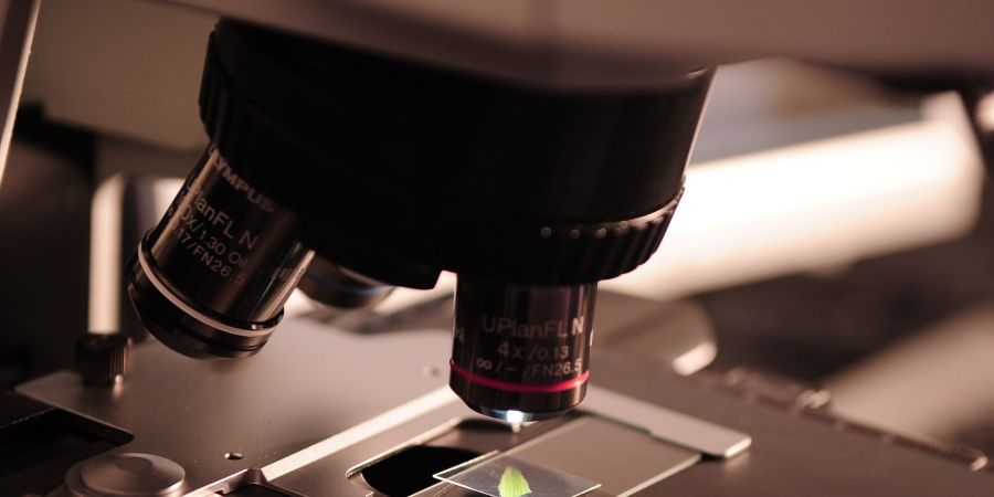
Microscopes are the important instrument which is used for the study of microbial structures .They are different kinds of microscopes are used for observing microbes and molecules.
Light microscopes uses glass lenses to bend and focus light rays to produce the enlarged images of small objects.It is classified as,
- Bright field microscopes is an ordinary microscopes used in all research labs.It forms a brighter image against a brightest background.Its components are ‘eyepiece or ocular lenses’ and the 'objective lenses'.Its magnifying lenses ranges from 10x to 100x .
- Dark field microscopes is used for observing unstained living cells organisms.It is used for revealing larger structure of eukaryotic cells and used to identify the thin and distinct shaped bacterial cells such as Treponema pallidum.
- Phase Contrast microscopes is for observing unpigmented cells.It converts the slight difference in refractive index and cell density into easily detected variations in light intensity and is an excellent way to observe living cells.
- Fluorescence microscopes exposes a specimen to ultraviolet rays and forms image of an object i.e,the fluorescent light is emitted very quickly to the excited molecule and the trapped energy returns to a more stable state
Staining of specimen
For observe the morphology of microbes in microscopy,we need to prepare the specimen Sample.The stained samples are reprenting the living cells as closely as possible.
Fixation is the process in which the internal and external structure of cells are fixed in position.A microorganism is usually killed and attached to microscope slide during fixation.It is classified as
- ‘Heat fixation’ is used to observe prokaryotes.It is done by heating the cells gently as a slide is passed through a flame.It preserves the morphology bit not structures within the cells.
- ‘Chemical fixation’ in which the chemical fixatives penetrate cells and react with cellular components, usually proteins and lipids to make them inactive,insoluble and immobile.Common fixative mixtures are ethanol,acetic acid,formaldehyde.
Staining are classified as
- Simple staining
- Differential staining
- Flagella staining.


