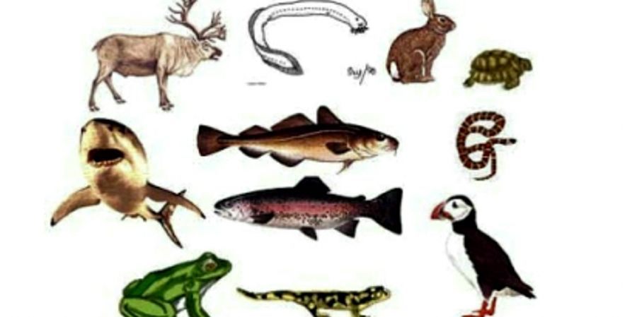

The animals which are characterised by the presence of a solid, unjointed, elastic, dorsal notochord; tubular, hollow nerve cord, situated dorsal to the notochord and pharyngeal gill slits are called chordate animals in the phylum Chordata. This phylum marks the climax of animal evolution. The chordates are the most familiar, most adaptable, most successful and most widely distributed animals. They exhibit great diversity of form, habitat and habits. There are some 65,000 known living chordates and most of them are free-living.
The phylum Chordata includes animals that can be grouped into two or sub-phyla: the Vertebrata (Craniata) and major subdivisions Protochordata (Acraniata).
Three Fundamental Chordate Characters :- All the chordates possess three outstanding unique characteristics at some stage in their life cycle. These three fundamental morphological features include:
1).Dorsal hollów or tubular nerve cord: The central nervous system of the chordates is located dorsally in the body. Nerve cord is tubular, hollow and longitudinal situated just above the notochord and extending lengthwise, in the body. The nerve cord or neural tube is derived from dorsal ectodermal neural plates of the embryo and encloses a cavity or canal called neurocoel. There are no ganglia. The nerve cord performs the functions like integration and co-ordination of the body activities. In vertebrates, the anterior part of the nerve cord is specialized into brain and protected in the cranium. The posterior of nerve cord is called spinal cord which is protected in vertebral column.
2). Longitudinal supporting rod-like notochord: It is also called chorda dorsalis. It is rod-like, flexible structure extending the length of the body. It is present beneath nerve cord and just above the digestive system. It is originated from the endodermal roof of the embryonic archenteron. It is formed by large vacuolated notochordal cells containing gelatinous matrix and surrounded by outer fibrous and an inner elastic sheath. It gives support to the body or act as internal skeleton. In vertebrates, it is replaced by vertebral column.
3). Pharyngeal gill slits: In all the chordates, at some stages of their life history, a series of paired lateral gill clefts or gill slits leading outward from the pharynx. They are also known by different names like pharyngeal clefts or pouches, branchial or visceral clefts. They are endodermal in origin. They are useful for water passage from pharynx to outside, thus, bathing the gills for respiration. The water current secondarily aids in filter feeding by capturing food particles in the pharynx. In protochordates
(e.g. Branchiostoma) and lower aquatic vertebrates the gill slits are functional throughout the life.
Visit my website biologylower45.blogspot.com


