 Heart is the main organ of circulatory system, a web of blood veins that circulates blood throughout the body.It also coordinates with other bodily systems to regulate blood pressure and heart rate. The heart functions is influenced by our family history, personal health history, and lifestyle.
Anatomy of heart:
The heart is a fist-sized organ that pumps blood throughout your body. It is the main component of the circulatory system.
The four major muscle-powered chambers that make up the heart are propelled by electrical impulses. Heart's activity is guided by brain and neurological system.
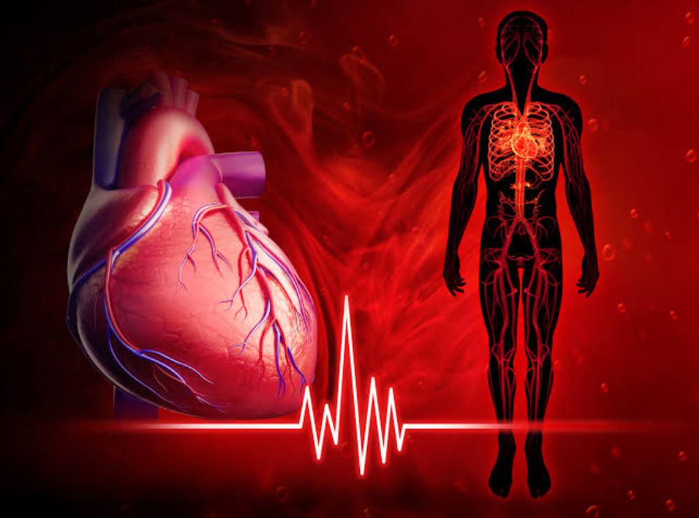
Location of the heart.
Heart is located in the front of chest. It is situated just left and slightly behind your sternum (breastbone). The rib cage guards the heart.
Purpose of the heart:
The major job of the heart is to circulate blood throughout your body.
1. Heart regulates the tempo and rhythm of our heartbeat.
2.Keeps our blood pressure steady.
How do your heart and other organs interact?
Heart regulates heart rate and other bodily processes in conjunction with other bodily systems. The fundamental systems are:
1.Nervous system aids in the regulation of your heart rate. When you are at rest or under stress, it transmits signals telling your heart to beat more quickly.
2. Endocrine system produces and releases hormones. Blood vessels are instructed to tighten or relax by these hormones, which has an impact on blood pressure. Thyroid gland's hormones can also instruct heart to beat more quickly or more slowly.
Outline view of heart:
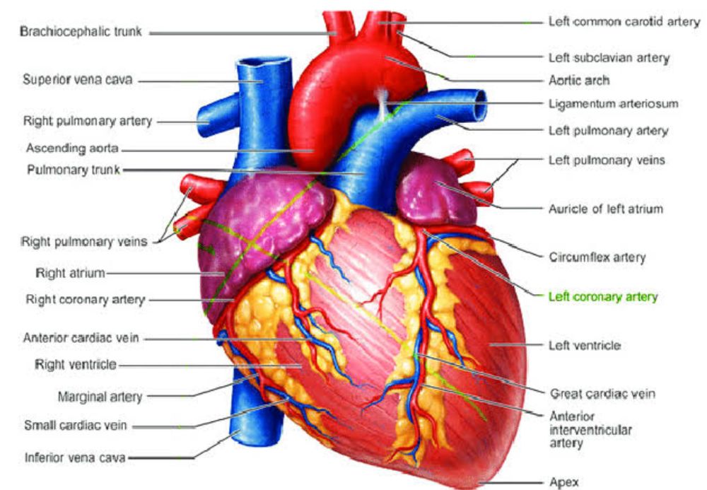
Borders:
Superior (atria, auricles), inferior (right and left ventricles), left (left auricle, left ventricle), right (right atrium) borders
Surfaces:
Sternocostal (right ventricle), diaphragmatic (mostly right ventricle, portion of left ventricle), pulmonary (cardiac impression) surfaces
Chambers:
Atria (left and right), ventricles (left and right)
Emerging/entering vessels:
1.Pulmonary veins (-> left atrium)
2. Superior and Inferior vena cavae (-> right atrium)
3. Aorta (left ventricle ->)
4. Pulmonary artery (right ventricle ->)
Valves:
1.Tricuspid valve
2.Pulmonary valve
3.Mitral valve
4.Aortic valve.
Detail view of heart structure:
Four chambers make up heart. The chambers resemble rooms in your heart's "home." These chambers are divided into upper and lower chambers as well as left and right chambers
The two upper chambers are called atria. They are right and left atrium.
The two lower heart chambers are called ventricles. They are left and right ventricles.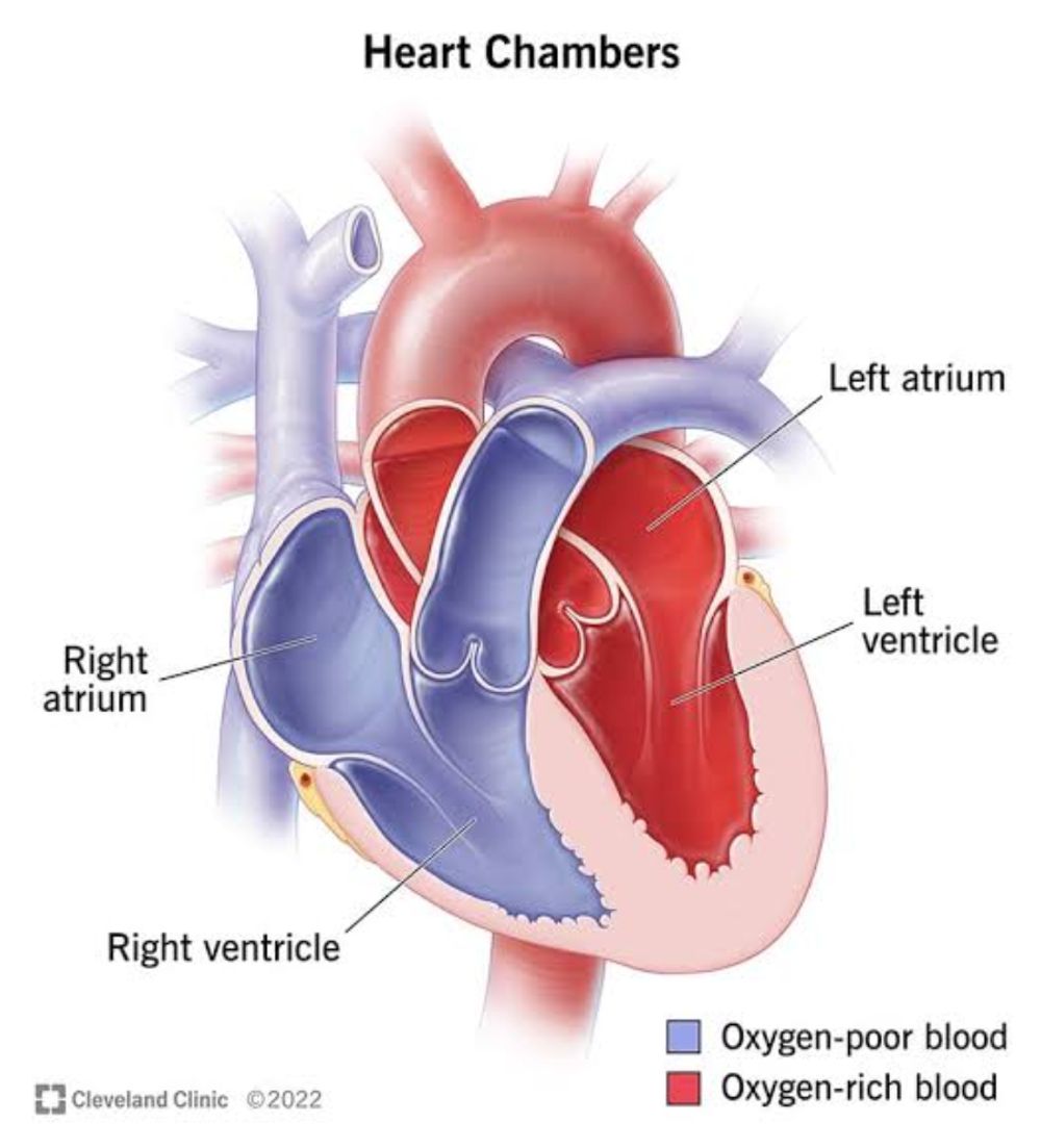
Heart valves are located in between the top and bottom chambers. Valves are present to ensure proper blood flow. heart valves open and close during blood circulation . Blood only flows in one way. .
1. Left ventricle and aorta are connected by an aortic valve (large artery that carries blood throughout your body).
2. Left atrium and left ventricle are connected via the mitral valve or bicuspid valve which is made up of two slender tissue.
3.Right ventricle and pulmonary arteries are connected by a pulmonary valve (arteries that carry blood to lungs).
4.Right atrium and right ventricle are connected by the tricuspid valve or right atrioventricular valve, which is made up of three tiny flaps of tissue.
Blood flow :
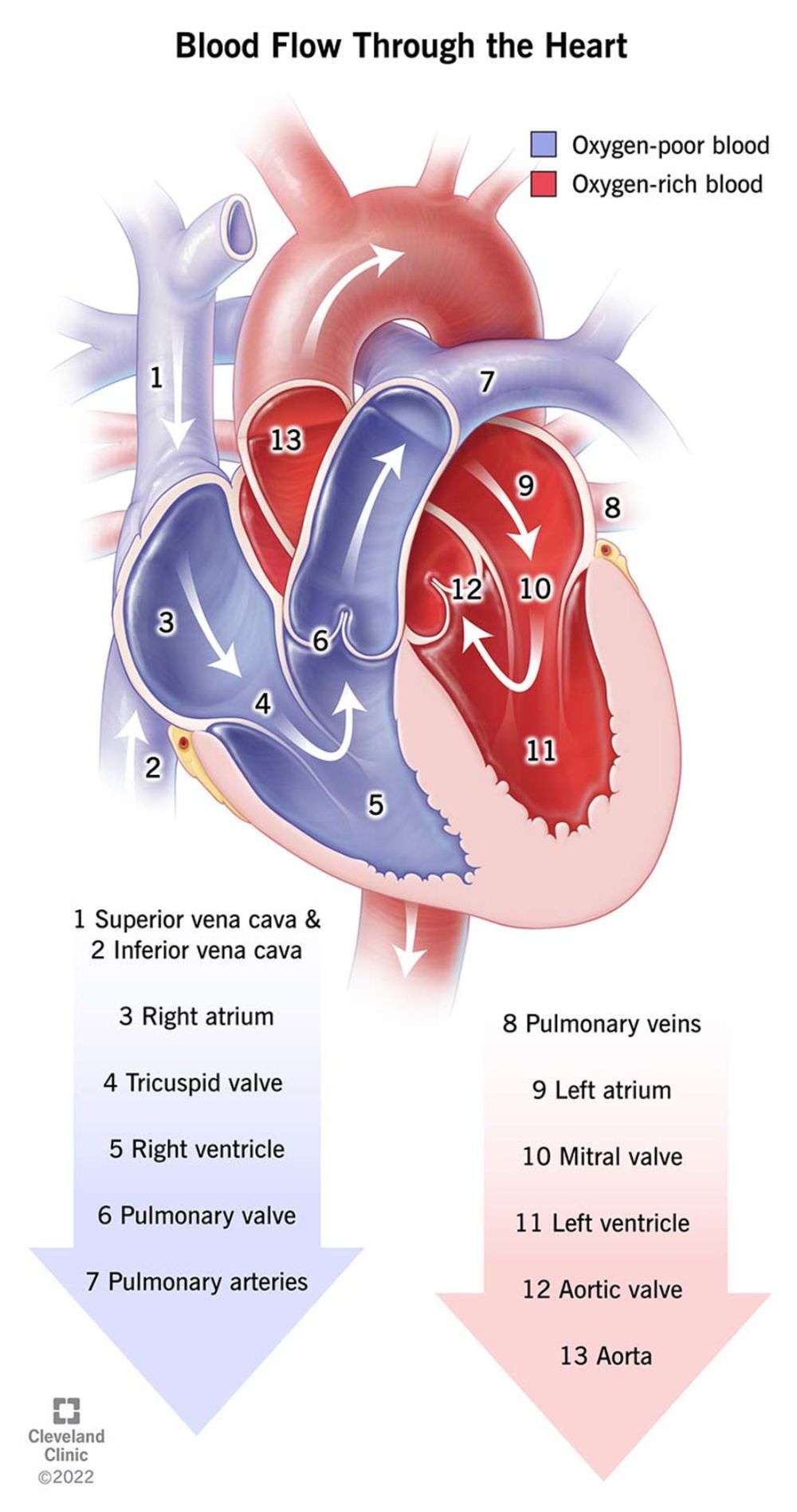
The heart's blood flow includes the following: