Published Sep 6, 2021
2 mins read
476 words
This blog has been marked as read.
Read more
Medical
Academics and Education
Science

Gram Staining -: Primary Test To Be Perform

Published Sep 6, 2021
2 mins read
476 words

                   Hello everyone!!!!

              Today I will tell you about one of the primary tests we learn in Microbiology Laboratory in college. 

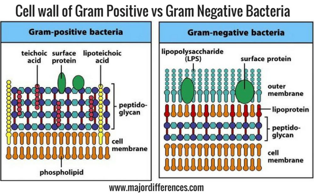
               We all are aware of basic cell structure. Outer layer of the cell is know as cell wall. Peptidogycan is major compound found in call wall which is basically composed of sugar and amino acid molecules. Thickness of the peptidoglycan in cell wall differentiate bacteria in two categories Gram Positive ( which has thick peptidoglycan layer about 50-90% of cell wall ) and Gram Negative ( which has thin peptigoglycan layer about 10% of cell wall ) as show in below picture .
   Mechanism of Staining -:

            Crystal violet , first dye used in this staining penetrates into cell wall of both Gram positive and negative cells.positive ions of the dye interacts with negative ions in the membrane and cell wall gets stain in violet in colour.After adding iodine ion in it Crystal Violet Iodine complex (CVI) get form in the wall.

            When decolorizer such as alcohol or acetone used in it, due multilayered nature of the peptidoglycan of Gram Positive cell,the CVI complex gets strapped and cannot remove by the addition of decolorizer. Hence Gram Positive Bacteria retain the stain and remain perple. Whereas in the case of Gram Negative bacteria, alcohol dissolves outer lipid area , leaving peptidoglycan layer exposed and it lead to removal of CVI complex making Gram Negative bacteria colourless. 

                     A counter stain, basic fuchsin, stains the Gram Negative cells pink, differentiate them from purple Gram Positive bacteria.This test is important in characterization and classification of bacteria.It is also used in screening of infectious agent in clinical specimens.

Lets go for actual procedure of the staining -:

  1. Take clean grease free glass slide.
  2. Put bacteria which in test tube in saline suspension on glass slide with the help of wire loop and dry the saline suspension with the help of burner. it is also called as heat fixing.
  3. Stain the slide with crystal violet for 1 min.Discard the stain and wash it gently.Don't wash it under forcefully running tap water.
  4. Flood the slide with Gram iodine for and let it react for 1 minute.Discard the iodine but do not  wash with water,
  5. Hold the slide in slanting position and allow 1-2 drops of alcohol and let it drain quickly.
  6. Wash the slide with water and them stain it with basic fucshin. let it react for 1 min.
  7. wash slide gently with water and it is ready to observe under 100x oil immersion lens light microscope.( put one drop of oil on slide and make sure your lens is getting immersed into oil.

We get to observe something like this under microscope -:
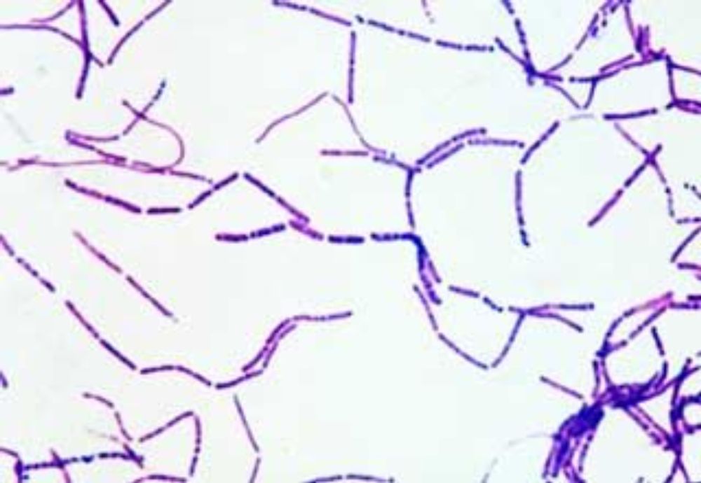
Bacillus Subtilis - Gram Positive and purple in color 
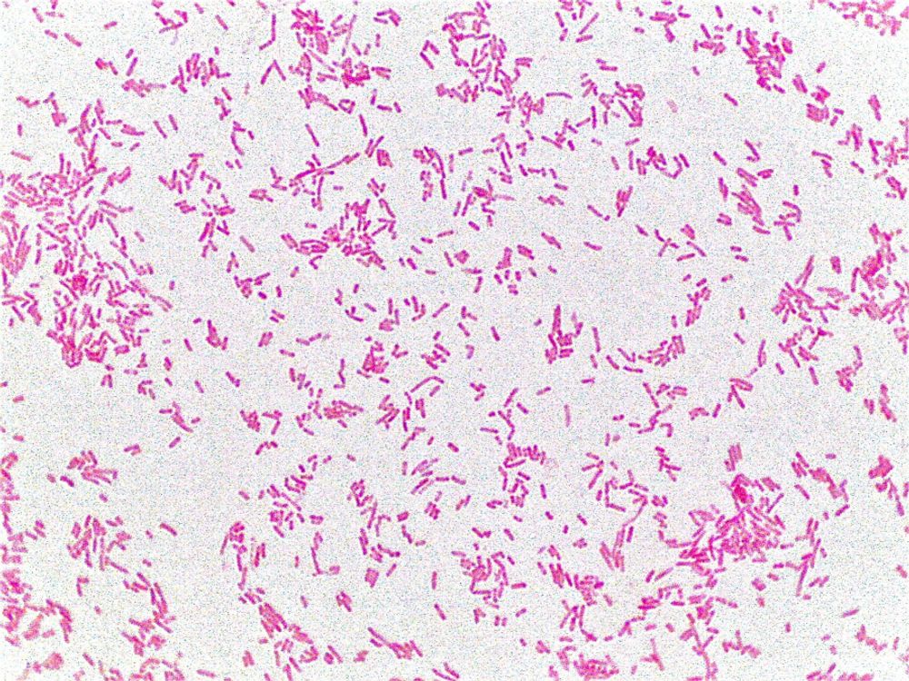
Escherichia Coli - Gram Negative and pink in color 

  Do like, comment and share.

Suggestions are always welcome.

#science
#microbiology
#staining
11
3
asadmirza1997 9/6/21, 8:00 AM
👍👍👍👍👍👍
niharika.kunder 9/16/21, 8:53 AM
informative
lokeshbhandari821 5/13/22, 1:26 PM
Nice Blog Pls read mine too

Candlemonk | Earn By Blogging | The Bloggers Social Network | Gamified Blogging Platform

Candlemonk is a reward-driven, gamified writing and blogging platform. Blog your ideas, thoughts, knowledge and stories. Candlemonk takes your words to a bigger audience around the globe, builds a follower base for you and aids in getting the recognition and appreciation you deserve. Monetize your words and earn from your passion to write.