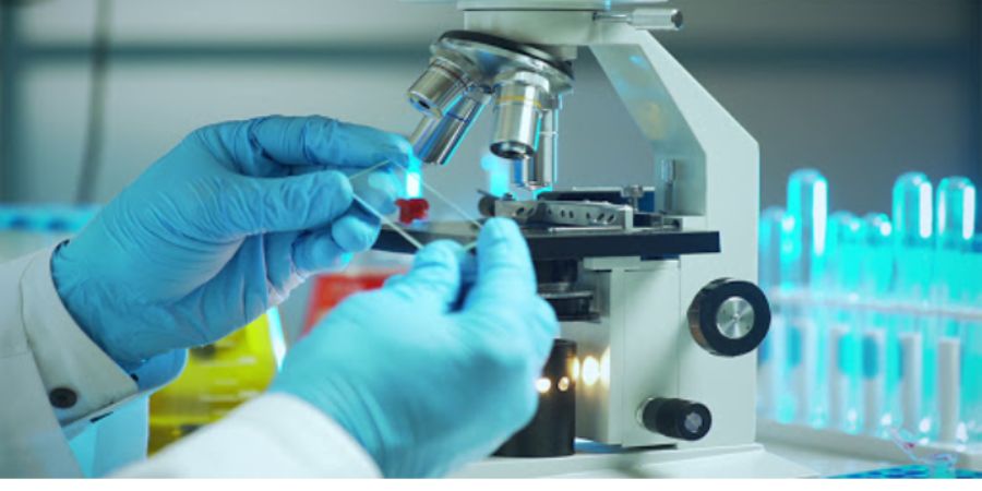

Dark field/Dark ground microscope:
Another method of improving the contrast is the dark field microscope in which reflected light is used instead of the transmitted light used in the ordinary microscope.The essential part of the dark field microscope is the dark field condenser with a central circular, which illuminate the object with a cone of light,with out letting any ray of light falls directly on the objective lens.light rays falling on the object are reflected or scattered on to the objective lens, with the result that the object appears self luminous against a dark background.The contrast gives an illusion of increased resolution,so that very slender organism such as spirochete,not visible under ordinary illumination,can be clearly seen under the dark microscope.
The resolving power of the light microscope is limited by the wavelength of light.In order to be seen and delineated,an object has to have a size of approximately half the wavelength of the light used.with visible light,using the best optical system,the limit of resolution is about 300 num.If light of shorter wavelength is employed,as in the ultraviolet microscopes, the resolving power can be proportionately extended.
Two specialized types of microscopeThe interference microscope which not only reveal cell organelles but also enable quantitative measurements of the chemical constituents of cells such as lipids, protein and nucleic acid and the polarisation microscope which enables the study of intracellular structure using differences in birefringence.
Electron microscope
In the electron microscope,a beam of light used in the optical microscope.The electron beam is focused by circular electromagnet, which are analogous to the lenses in the light microscope.The object which is held in the path of the beam scatters the electron and produces an image which is focused on a fluorescent viewing screen.As the wavelength of electron used is approximately 0.005 nm,as compared to 500 nm with visible light,the resolving power of the electron microscope should be theoretically 100,000 times that light microscope but in practice, the resolving power is about 0.1nm.
The technique of shadow casting with vaporised heavy metals has made picture with good contrast and three dimensional effect possible.Another valuable techniques in studying fine structure is negative staining with phosphotungstic acid.gas molecules scatter electron,and it is therefore necessary to examine the object in a vacuum, hence,only dead and dried object can be examined in the electron microscope.


