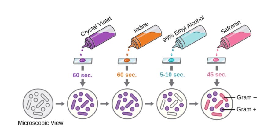

Today my vlog is related to Principle of gram staining
The gram staining property is determined by the cell Wall of bacterium.Gram negative bacteria contain a thin pepti layer above which a lipid rich outer membrane is present.On
treatment with a decolourizer, the lipid layer of the cell Wall is destroyed exposing the thin peptidoglycon layer.The dye iodine complex easily comes out of this thin layer of glycon and hence gets decolourized.on the other hand the gram positive bacteria contain only a thick peptidoglycon layer as the cell Wall.They are not affected by the decolourizer.How ever the thick peptido layer get dehydrated there by closing the pores present in them.Thus the dye iodine complex is retained even after decolour.Acid fast staining there are certain bacteria whose cell Wall is rich in mycolic acid and hence cannot penetrate such waxy cell Wall.one such example of bacteria is mycobacterium species acid fast staining techniques also come under the differential staining method.They differentiate bacteria as acid fast and non acid fast bacteria.
The routinely used methods of acid fast staining is the ziehl neelsan method.It has three steps
Primary staining, Decolourizer, counter stain.
Step one the primary stain used is strong carbol fuchsin which phenol the phenol help in the penetration of Stain into the cell Wall.the penetration can further be assisted by heating the stain which loosen the cell Wall.
Step two Decolourizer the used is either sulphuric acid or alcoholic hydrochloric acid.All non acid fast organisms will lose the primary Stain on treatment with acid decolourizer where as the acid fast organisms retain the primary stain.
Step three Counter stain methylene blue is added as counter stain the decolourized bacteria will take up the counter Stain and appear blue in colour.The acid fast bacteria do not take the counter Stain and hence appear pink in colour.
Acid-fast bacillus staining principle Since acid-fast bacilli contain the cell wall of acid-fast bacilli,
retains the primary staining and appears to be stained pinkish blue.
Apart from the staining methods described here, there are many special staining methods to show other morphological features that help identify the bacterium, such as flagellar spore capsules. Thank you very much Determination of sodium
Na+ has been measured in various ways, including chemical methods, flame emission spectrophotometry, atomic absorption spectrophotometry (AAS), and ISEs.
Chemical methods are outdated because of large sample volume requirements and lack of precision.
ISEs are the most routinely used method in clinical laboratories.


